


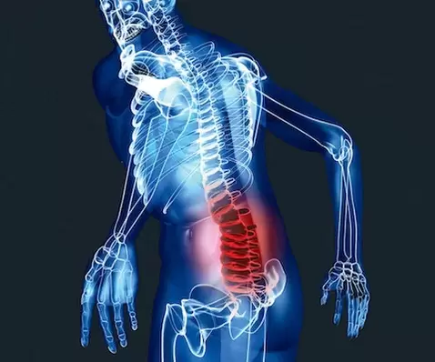
Osteochondrosis of the lumbar spine: symptoms and treatment
The causes of osteochondrosis of the lumbar spine are not well understood. The greatest importance is given to hereditary predisposition, age-related changes in the intervertebral discs. Pain can be provoked by uncomfortable movement, prolonged forced position, lifting and carrying heavy loads, sports overload, overweight.
Depending on the duration, there are acute pains lasting up to 4 weeks, subacute (from 4 to 12 weeks) and chronic (lasting more than 12 weeks).
Neurological complications of osteochondrosis of the lumbar spine:
Lumbago (lower back pain). Acute pain in the lumbar region begins suddenly, provoked by minimal movements in the back. Movement in the lumbar spine is sharply limited, there is compensatory scoliosis. Paravertebral muscles with "stone" density. The duration of lumbago with adequate treatment and immobilization of the lumbar spine is no more than 7-10 days.
Lumbodynia (back pain).Patients complain of moderate pain in the lumbar region, which increases when moving or in a certain position, discomfort when standing or sitting for a long time. The onset is usually gradual. Clinically, limited mobility in the lumbar spine, tension and soreness of the paravertebral muscles is often determined. In most cases, the pain subsides within 2-3 weeks, but if left untreated, it can become chronic.
Lumbosciatica (pain in the lower back radiating to the leg). In the lumbar region, movements are limited, the paravertebral muscles are tense and painful on palpation.
In piriformis syndrome, the sciatic nerve is compressed, causing paresthesias and numbness in the leg and foot. Positive Lasegue syndrome. But there are no signs of radicular syndrome.
Disc herniation with radicular syndrome or radiculopathy. Compression of the root is accompanied by a shooting, burning pain in the leg. The pain intensifies when moving, when coughing, accompanied by tingling at the root, muscle weakness and loss of reflexes. Positive stress symptoms.
In the lumbar region, the greatest load falls on the lower part, therefore the L5 and S1 roots are most often involved in the pathological process. Each root has its own area of distribution of pain and numbness in the limbs.
Radicular syndromes are detected by a neurologist during an objective examination.
Vascular-radicular conflict. Paralyzing sciatica syndrome occurs when blood circulation is impaired in the L5 and less commonly S1 radicular artery. Radiculoischemia at other levels is diagnosed extremely rarely.
With uncomfortable movement or heavy lifting, sharp pain in the back develops with irradiation along the course of the sciatic nerve. Then there is paresis or paralysis of the extensors of the foot and toes with "hitting" of the foot when walking (stepping). The patient, while walking, raises his leg high, throws it forward and at the same time hits his toe on the floor.
In most cases, paresis safely regresses within a few weeks.
Violation of blood supply to the spinal cord and cauda equina. In spinal stenosis, several spinal nerve roots (cauda equina) are affected. The pain at rest is minor, but there is an intermittent limping syndrome when walking. Pain when walking spreads along the roots from the lower back to the legs, accompanied by weakness, paresthesia and numbness of the legs, disappears after rest or when the torso is bent forward.
Acute disturbance of spinal blood circulation is the most serious complication of lumbar osteochondrosis. Acutely develops lower paraparesis or plegia. Weakness in the legs is accompanied by numbness of the lower limbs, dysfunction of the pelvic organs.
Review of patients with osteochondrosis of the lumbar spine.
Of great importance is the analysis of complaints and history to rule out serious pathology. A neurological examination is performed to exclude damage to the roots and spinal cord. Manual examination allows you to determine the source of pain, limitation of mobility, muscle spasm.
Additional research methods are indicated when specific back pain is suspected.
An X-ray of the lumbar spine is prescribed to rule out tumors, spinal injuries, spondylolisthesis. X-ray signs of osteochondrosis have no clinical value, since all adults and elderly people have them. Functional X-rays are taken to look for spinal instability. Photographs are taken in a position of ultimate flexion and extension.
For radicular or spinal symptoms, an MRI or CT scan of the lumbar spine is indicated. On MRI, disc herniations and the spinal cord are better seen, and on CT, bone structures are better seen. The clinical level of the lesion and the MRI findings must match, as a herniated disc detected on MRI is not always the cause of the pain.
Electroneuromyography (ENMG) is sometimes prescribed for neurological deficits to clarify the diagnosis.
If somatic pathology is suspected, a thorough clinical examination is performed.
Osteochondrosis of the lumbar spine, treatment.
When the first signs of discomfort in the lumbar spine appear, regular gymnastics to strengthen the muscle corset, swimming courses and massage are indicated.
Treatment of lumbar osteochondrosis is divided into 3 periods: treatment of acute, subacute and chronic period.
In the acute period, the main task is to relieve the pain syndrome as early as possible and restore the patient's quality of life. In the presence of intense pain, immobilization of the lumbar spine with a special antiradiculitis corset for 2-3 weeks is indicated. Bed rest should not last more than 2-3 days. In many patients, it is possible to increase the pain syndrome against the background of the expansion of the motor mode. The patient should not limit himself to reasonable physical activity.
Of the non-drug methods of therapy, interstitial electrical stimulation, acupuncture, hirudotherapy and massage are effective. It is possible to use manual therapy, but only in competent hands.
Medical treatment. Nonsteroidal anti-inflammatory drugs are indicated for acute pain. In combination with anti-inflammatory drugs, muscle relaxants can be prescribed in a short course.
In osteochondrosis of the lumbar spine, therapeutic blockades with local anesthetics, non-steroidal anti-inflammatory drugs and corticosteroids are effective. Medicinal mixtures are applied as close as possible to the focus of pain (in the affected muscles, the starting points of the roots).
In radiculopathy with the presence of neuropathic pain, anti-inflammatory drugs are ineffective, in this case antidepressants, anticonvulsants and a special therapeutic patch are prescribed.
In case of paresis, numbness, vascular preparations, vitamins of group B are prescribed.
For prolonged myofascial pain, the introduction of nonsteroidal anti-inflammatory drugs into trigger points, muscle relaxants, acupuncture, and postisometric relaxation is effective.
For chronic pain, antidepressants, exercise therapy, and other non-pharmacological treatments are the first line of treatment.
For stenosis of the spinal canal, weight loss, wearing a corset, NSAIDs and various venotonics are indicated.
Surgical treatment is performed with paralyzing sciatica (during the first three days) and cauda equina syndrome (paresis of the limbs, impaired sensitivity, urinary and fecal incontinence).
Prevention of lumbar osteochondrosis
Preventionosteochondrosis of the lumbar spinereduced to avoiding long, uncomfortable positions, excessive loads. It is important to properly equip your workplace, to alternate periods of work and rest. Wear a restraint belt for physical overload. Do exercises to strengthen your back muscles.